


All product descriptions and articles provided on this website are intended strictly for informational and educational purposes. Our products are designed exclusively for in-vitro research (i.e., experiments conducted outside of a living organism, typically in glassware such as test tubes or petri dishes). These compounds are not approved by the FDA for use in humans or animals. They are not medications, nor are they intended to diagnose, treat, prevent, or cure any disease or medical condition. Any bodily administration-human or animal-is strictly prohibited by law. Our products are not for human consumption under any circumstances.
What Evidence Supports NAD+ Boosting Peptides in Guarding Neurons During Stroke?
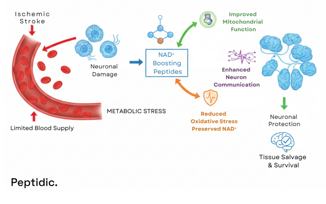
Peptide-based NAD⁺ enhancers show strong potential for protecting brain cells when the blood supply is limited during stroke. They help maintain stable cellular energy production and promote healthier mitochondrial function. These peptides also support everyday neuron communication while reducing the harmful metabolic stress that drives cell death. Studies in stroke models consistently link NAD⁺ preservation to reduced tissue damage and improved chances of neuronal survival.
Peptidic delivers high-quality research peptides trusted for precision in advanced neuroscience studies. Our knowledgeable team supports researchers in solving technical challenges, accelerating experimentation, and uncovering new NAD⁺ mechanisms in stroke research. Partner with Peptidic and advance your discoveries with products and expertise designed to drive meaningful scientific progress.
How Does NAD⁺ Control Neuronal Survival During Ischemia?
NAD⁺ controls neuronal survival during ischemia by sustaining energy production when blood flow decreases. Additionally, research from the University of Oxford [1] indicates that NAD⁺ depletion leads to the collapse of ATP, damages mitochondria, and increases oxidative stress. Therefore, preserving NAD⁺ delays this injury cascade and helps neurons withstand ischemic conditions more effectively.
These key biological actions help sustain neurons under stress:
- Prevents excessive NAD⁺ depletion linked to rapid energy collapse
- Maintains mitochondrial efficiency to reduce oxidative stress
- Supports essential synaptic communication during blood flow interruption
Therefore, protecting NAD⁺ during a stroke preserves neuronal stability and reduces the severity of injury. It also helps control metabolic failure, which improves overall brain resilience in ischemic events.
How Do NAD⁺ Peptides Deliver Neuroprotection During Ischemia?
NAD⁺ peptides deliver neuroprotection during ischemia by sustaining cellular energy when blood flow drops. Moreover, research from Harvard DASH[2] shows they enhance sirtuin signaling and reinforce NAD⁺ recycling. Therefore, they help prevent rapid mitochondrial decline that would otherwise lead to neuronal cell death.
These key pathways explain their protective impact:
1. Sirtuin Activation
NAD⁺ peptides boost sirtuin enzyme activity that regulates mitochondrial performance and cellular defense systems. This reduces oxidative stress, strengthens neuronal function, and helps brain cells resist the early damage triggered by ischemia.
2. Enhanced NAD⁺ Salvage Pathway
These peptides support NAMPT and NMNAT pathways to rebuild NAD⁺, even after oxygen loss. Sustained NAD⁺ improves ATP production and lowers neuron vulnerability to ischemic metabolic failure.
3. Mitochondrial and Axonal Stability
By maintaining NAD⁺ availability, peptide interventions preserve mitochondrial respiration and protect axonal structure. Neurons remain functional longer and avoid catastrophic energy collapse during stroke events.

What Research Confirms the Neuroprotective Effects of NAD⁺ Peptides in Stroke Models?
Experimental research confirms that NAD⁺-focused peptides help protect neurons during ischemic stroke by maintaining brain energy levels when blood flow is disrupted. For example, Johns Hopkins' [3] study shows that excessive PARP-2 activation increases infarct size and accelerates the damaging release of AIF. However, NAD⁺ preservation prevents this rapid mitochondrial breakdown and supports cellular structure. Therefore, stabilizing NAD⁺ directly limits the early chain of metabolic failure seen in stroke.
Additionally, the restoration of NAD⁺ in the brain further strengthens the scientific foundation for peptide-based interventions against ischemia. University of Memphis[4] study found that oral NMN raised brain NAD⁺ by over 40 percent in mice. This boosted mitochondrial respiration and reduced cell loss during ischemic stress. Consequently, these results highlight how elevating NAD⁺ improves neuronal survival in controlled stroke research.
How Do Sirtuins and PARP Balance Support Neuronal Survival in Stroke?
Sirtuins and PARP balance support neuronal survival in stroke by maintaining NAD⁺ availability and protecting mitochondrial energy during reduced blood flow. Moreover, when sirtuins function properly and PARP overactivation is limited, oxidative stress remains low, and neurons avoid energy failure. Therefore, this balance leads to stronger neuronal survival outcomes in preclinical stroke studies.
Together, these processes strengthen neuronal resistance to ischemia:
- Sirtuins enhance mitochondrial defense: activate stress-response and DNA-repair enzymes using NAD⁺, lowering oxidative damage and helping neurons maintain energy during reduced blood flow.
- Controlled PARP activity preserves NAD⁺ supply: limits excessive DNA-repair demands that rapidly consume NAD⁺, keeping ATP production stable and slowing neuron death under ischemic stress.
- Balanced signaling protects neural communication: maintains mitochondrial structure and axonal integrity, allowing neurons to continue sending signals even when oxygen and nutrients are severely restricted.
Empower advanced ischemia research with NAD⁺ supporting peptide solutions from Peptidic
Researchers working on neuroprotection in ischemia models need reliable materials to accurately investigate NAD⁺-linked mechanisms. They depend on high-purity peptides, consistent performance, and uninterrupted supply. However, delays or batch variations can disrupt experiments and slow scientific progress. Therefore, ensuring dependable resources is essential for producing clear and trustworthy stroke-model results.
Peptidic delivers high-quality peptides such as NAD⁺ that support strong reproducibility in neurological studies. Our team works closely with scientists to simplify workflows and reduce technical setbacks. Additionally, we prioritize product reliability to help protect data quality. Contact Peptidic to accelerate innovative discoveries in ischemia and stroke-model research.

FAQs
How do NAD⁺ peptides support ischemia research?
NAD⁺ peptides support ischemia research by helping neurons maintain energy during blood flow reduction. Moreover, they stabilize mitochondria and reduce oxidative stress. Therefore, they enable clearer evaluation of neuroprotective effects in stroke-model experiments.
Why do researchers measure NAD⁺ levels in stroke models?
Researchers measure NAD⁺ levels to track metabolic failure during ischemia. Additionally, reduced NAD⁺ indicates mitochondrial stress and progressing neuronal injury. Consequently, this measurement validates the effectiveness of experimental neuroprotective strategies.
Why is peptide-driven NAD⁺ enhancement valuable in studies?
Peptide-driven NAD⁺ enhancement improves mitochondrial performance and cellular defense pathways under ischemic stress. Moreover, it strengthens neuronal survival outcomes. Therefore, it provides measurable advantages when analyzing stroke-related injury mechanisms.
How does PARP activity affect NAD⁺ during ischemia research?
PARP activity affects NAD⁺ by consuming it during DNA repair, reducing available energy in ischemia models. Moreover, excessive activation rapidly depletes NAD⁺ and worsens metabolic stress. Therefore, regulating PARP is essential for studying ischemic neuronal injury.

