


All product descriptions and articles provided on this website are intended strictly for informational and educational purposes. Our products are designed exclusively for in-vitro research (i.e., experiments conducted outside of a living organism, typically in glassware such as test tubes or petri dishes). These compounds are not approved by the FDA for use in humans or animals. They are not medications, nor are they intended to diagnose, treat, prevent, or cure any disease or medical condition. Any bodily administration-human or animal-is strictly prohibited by law. Our products are not for human consumption under any circumstances.
How Does NAD+ Deficiency Affect the Progression of Parkinson’s Disease?
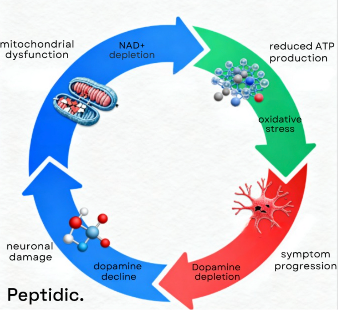
Parkinson’s disease (PD) is among the fastest-growing neurodegenerative disorders, projected to affect over 25 million people[1] by 2050. Beyond aging, research shows that mitochondrial dysfunction and declining NAD⁺ levels are key triggers of neuronal energy loss and oxidative stress. These metabolic failures accelerate dopamine neuron degeneration, highlighting NAD⁺ restoration as a promising approach to slow or modify Parkinson’s disease progression.
Peptidic is advancing the frontiers of neurodegenerative research by delivering ultra-pure, research-grade NAD⁺ and metabolic compounds engineered for scientific precision and reproducibility. Our formulations empower researchers to investigate the molecular effects of NAD⁺ restoration on neuronal survival, mitochondrial efficiency, and energy metabolism in Parkinson’s disease and related disorders, driving breakthroughs in translational and therapeutic discovery.
How Does NAD⁺ Support Neuronal and Mitochondrial Function?
NAD⁺ supports neuronal and mitochondrial function by powering cellular energy metabolism, stabilizing neuronal activity, and protecting against oxidative stress. It is essential for maintaining brain vitality and preventing the cellular decline associated with Parkinson’s disease.
Key functions explain their essential neurological role:
- Fuels ATP production[2] for neuronal energy
- Aids DNA repair and genomic stability
- Regulates calcium and signaling balance
When NAD⁺ levels decrease, mitochondrial efficiency declines, resulting in reduced ATP synthesis, impaired neurotransmitter release, and weakened synaptic communication. This metabolic disruption accelerates neuronal degeneration and amplifies Parkinson’s-related motor and cognitive symptoms.
Why Do NAD⁺ Levels Decline in Parkinson’s Disease?
NAD⁺ levels decline in Parkinson’s disease due to mitochondrial malfunction, oxidative damage, and disrupted biosynthetic pathways[3] that impair cellular energy balance. Genetic and environmental factors further block NAD⁺ regeneration, intensifying neuronal energy loss and accelerating neurodegeneration.
Key Biological Mechanisms Behind NAD⁺ Decline
- Impaired Electron Transport
Mitochondrial damage reduces NADH oxidation, limiting ATP synthesis and depriving neurons of essential energy.
- Oxidative Stress
Excess reactive oxygen species (ROS) harm mitochondrial DNA and enzymes, compounding cellular energy deficits.
- Genetic Mutations
Alterations in parkin and PINK1 genes disrupt NAD⁺ synthesis and recycling, weakening mitochondrial maintenance and neuronal survival.

What Research Links NAD⁺ Deficiency to Parkinson’s Disease Progression?
Multiple studies have revealed[4] a strong connection between declining NAD⁺ levels and the progression of Parkinson’s disease. Patients consistently show significant reductions in NAD⁺ and ATPmax levels, indicating widespread mitochondrial dysfunction. This energy imbalance weakens neuronal resilience and accelerates the loss of dopaminergic neurons, ultimately contributing to greater motor impairment and cognitive decline.
Clinical investigations further show that NAD⁺ depletion leads to fatigue, muscular weakness, tremors, and bradykinesia, hallmark symptoms of advanced Parkinson’s disease. Importantly, controlled trials indicate[5] that restoring NAD⁺ enhances mitochondrial efficiency, strengthens neuronal energy metabolism, and improves motor performance, highlighting its dual role as a therapeutic target and a potential biomarker for disease monitoring.
How Does NAD⁺ Deficiency Affect Neuronal and Muscular Performance?
NAD⁺ deficiency disrupts both neuronal and muscular systems by impairing mitochondrial energy production and weakening neurotransmission. The resulting energy shortfall reduces cellular vitality, leading to progressive muscle fatigue, neuronal apoptosis, and overall neurological decline characteristic of Parkinson’s disease.
Below are the major outcomes of reduced NAD⁺ levels.
Diminished Mitochondrial Function
Insufficient NAD⁺ limits ATP synthesis, depriving neurons and muscles of the energy required for optimal performance.
Decline in Muscular Endurance
Lower oxidative metabolism leads to early fatigue and reduced strength, significantly impacting mobility and coordination.
Accelerated Neuronal Degeneration
Energy-deprived neurons undergo programmed cell death, intensifying motor dysfunction and cognitive impairment.
Using ³¹P magnetic resonance spectroscopy[6], researchers have observed significant decreases in ATPmax among Parkinson’s patients, confirming that mitochondrial failure underlies much of the disease’s progression. Early restoration of NAD⁺ levels may, therefore, help preserve neuronal integrity and delay functional deterioration.
Advancing the Future of Parkinson’s Research with Peptidic’s High-Precision NAD⁺ Solutions
Parkinson’s research faces persistent challenges in tackling mitochondrial dysfunction, metabolic decline, and cellular energy imbalance, which are key drivers of neurodegeneration. Moreover, many therapeutic studies encounter issues with inconsistent compound quality and poor reproducibility. As a result, limited bioavailability further slows progress and complicates accurate interpretation of experimental outcomes.
Peptidic overcomes these limitations by providing research-grade NAD⁺ solutions engineered for unmatched purity, stability, and experimental precision. Our formulations are optimized for cellular and molecular research, enabling reproducible, high-fidelity results in studies of mitochondrial function and neuronal resilience. Contact us today to collaborate with Peptidic and advance the scientific understanding of Parkinson’s disease.

FAQs
What causes NAD⁺ loss in Parkinson’s?
NAD⁺ loss results from mitochondrial dysfunction, oxidative stress, and genetic mutations like parkin and PINK1. These factors disrupt NAD⁺ recycling and energy metabolism, reducing cellular resilience and accelerating neuronal degeneration in Parkinson’s disease.
How is NAD⁺ measured in research?
Researchers measure NAD⁺ using ³¹P magnetic resonance spectroscopy (MRS) and biochemical assays. These methods accurately assess NAD⁺ concentration, mitochondrial performance, and ATP production, offering vital insights into cellular metabolism and disease progression in both experimental and clinical Parkinson’s studies.
Why is NAD⁺ a biomarker?
NAD⁺ serves as a biomarker because it reflects cellular energy status and mitochondrial efficiency. Monitoring its levels helps detect early metabolic dysfunction, evaluate treatment response, and track Parkinson’s progression, making it a valuable marker in neurodegenerative research.
Why use research-grade NAD⁺?
Research-grade NAD⁺ ensures consistency, purity, and reproducibility across experiments. It eliminates variability caused by low-quality compounds, allowing scientists to produce reliable data on metabolic restoration, neuronal protection, and therapeutic mechanisms in Parkinson’s disease research.
References
6. Payne, T., Burgess, T., Bradley, S., Roscoe, S., Sassani, M., Dunning, M. J., Hernandez, D., Scholz, S., McNeill, A., Taylor, R., Su, L., Wilkinson, I., Jenkins, T., Mortiboys, H., & Bandmann, O. (2024). Multimodal assessment of mitochondrial function in Parkinson’s disease. Brain, 147(1), 267–280. https://doi.org/10.1093/brain/awad364

